Spinal Cord Neurons Responsible for Locomotor Behavior Identified
Walking is an essential behavior for animal survival and important to move around in humans. It has long been established that neurons responsible for such behavior reside within the spinal cord. Although the central commands to initiate or stop walking originate from different brain regions, it is neurons within the spinal cord that execute these commands. However, whether a single neuronal type is responsible for orchestrating such complex behavior has remained elusive until recently.
Using mice as the experimental paradigm, George Mentis, an associate professor in pathology and cell biology (in Neurology), and his team has recently reported at Cell, that a select type of neurons, known as ventral spinocerebellar tract neurons – or simply, VSCTs – are essential for such behavior.
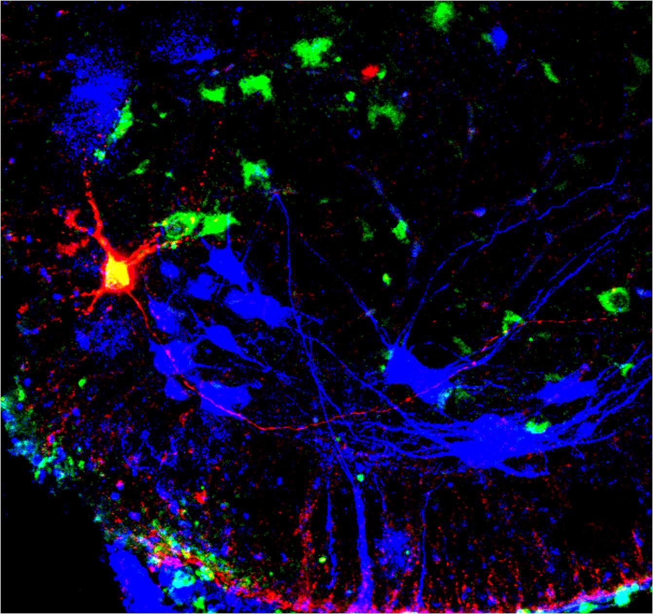
Confocal image from a mouse spinal cord, showing motor neurons (blue), ventral spinocerebellar tract (VSCT) neurons (green). A single VSCT neuron was recorded and marked intracellularly with a red dye.
VSCTs were discovered in the 1940s by the renowned neuroscientist Sherrington, and in the early 1970s Lundberg proposed that their main function was to relay messages about neuronal activity from the spinal cord to the cerebellum. However, the new study reports that instead they control locomotor behavior both during development and in adulthood.
Joshua Chalif, a highly talented MD/PhD student and first author of the study, together with Dr. Maria Martinez-Silva, an experienced and gifted postdoctoral research scientist in the Mentis Lab utilized a multitude of experimental approaches to dissect the function of VSCTs in mice. They utilized state-of-the-art mouse optogenetics, employing LED light to regulate certain proteins that were expressed selectively in VSCTs, introduced by viral means, to either activate or suppress the VSCTs’ neuronal activity. Leveraging on the ability of intact spinal cords from newborn mice to function in isolation within a dish (ex vivo), they demonstrated that activation of VSCTs by light, induced locomotor behavior. When VSCT activity was suppressed, ongoing locomotor behavior was halted.
The team explored further the function of these neurons in freely moving adult mice (in vivo), using chemogenetics techniques. Chemogenetics is a process by which a chemical compound is used to activate synthetic ligands expressed artificially and selectively in these neurons, controlling their activity. Freely moving adult mice stopped moving when the activity of VSCT neurons was suppressed by injecting the drug CNO (clozapine), in mice in which inhibitory ligands were selectively expressed in VSCTs. Similarly, locomotor behavior was also tested by the ability of mice to swim. Mice were unable to swim and simply floated in the water when VSCTs were silenced.
In all of these experiments, Dr. Mentis and his team demonstrated that VSCTs alone were both necessary and sufficient for controlling locomotor activity—activating them was enough to induce activity while suppressing them was enough to stop it.
“These findings were a huge surprise,” Dr. Mentis states. “One of the key discoveries in our study was that apart from their connection to the cerebellum, these neurons make connections with other spinal neurons that are also involved in locomotor behavior via the VSCTs’ axon collaterals.”
For their next steps, the Mentis Lab plans to identify and map precisely the neuronal circuits that VSCTs make with motor neurons and other spinal neurons. They also aim to identify select genetic markers and uncover potential subpopulations of VSCTs and explore their role in different modes of locomotion. Finally, they plan to explore how the function of VSCTs is altered in the context of pathology and neurodegenerative diseases.
Additional information
The paper is titled “Control of mammalian locomotion by ventral spinocerebellar tract neurons”; (2022) Cell, 185:328-344.
All authors: Joshua I. Chalif (Columbia), María de Lourdes Martínez-Silva (Columbia), John G. Pagiazitis (Columbia), Andrew J. Murray (University College London), and George Z. Mentis (Columbia).
The research was supported by the NIH (grants R01-NS078375, R01-AA027079, R21-NS079981 to G.Z.M. and F30NS098551 to J.I.C.), the SMA Foundation, and Project-ALS.